The Mechanisms of Connective Tissue and the Phases of Healing Following Whiplash
By Matthew J. DeGaetano, DC and Adam Shafran, DC
I was recently retained as an expert for a personal-injury case, in which a vehicle was struck from the rear end and forced the victim’s car to hit the vehicle in front of him. This occurred while the vehicle was stopped at an intersection; this collision would be defined as a double impact.
Here is the image of his vehicle:


 On the surface, the impact looks minor. But this was a hummer after all.
While the property damage was considered “minor”, the victim a 47 year old male (Front seat passenger) was ejected from his seat and caused his head strike the windshield thus, throwing back into the drivers seat.
The peer review doctor stated that this victim should have only received limited treatment because the patient acute phase of injury repair was 48 hours and did not require more treatment, he recommended 6 treatments max.
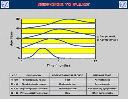
RANGES OF RECOVERY ON A 12 MONTH SCALE
 The victim sustained a C5-C6 disc herniation; this would obviously require more than 6 visits. Flattening of the spine with compression would occur when the victim slammed back into the seat.
Function and Properties of Discs
Cervical discs have the highest proportional height and their nuclei have the greatest capacity to swell. This allows greater range of motion and shock attenuation. The outer portion of the anulus is attached by Sharpey’s fibers which may be torn in cervical acceleration/deceleration (CAD) injuries. Discs, longitudinal ligaments and other adjacent paraspinal soft tissues can be torn during whiplash trauma (1-19,1137,1138).
Discs, like all soft tissues in the body, are viscoelastic structures. They display properties of anisotropy, creep and hysteresis.
- Anisotropy—the disc’s strength and stiffness vary depending on the type and direction of forces applied.
- Creep—the disc deforms gradually under a constant load.
- Hysteresis—the tissue loses energy after repetitive loading and unloading cycles.
The fluid dynamics of the nucleus in a healthy disc allows for an even distribution of load over the end plate (20).
Not only was the peer review wrong, crash tests at Spine Research Institute of San Diego, as well as those of others, have demonstrated a strong vertical acceleration of the lumbar and thoracic spine during the initial phase of a side-impact crash test (i.e., the so-called vertical ramping effect). This produces a large axial compressive force in the lumbar region. This would produce a horizontal (extensile) line of force, and would coincide with ramping and compression, thus creating injury to the lumbar facet joints and soft tissue.
Stress Characteristics of Ligaments

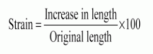
Understanding strain characteristics of ligament is crucial for the understanding of the types of injuries seen clinically (25). Tensile failure loads and maximal deflection at failure of all cervical ligaments is presented in my textbook [pp 13-14] (1402). In isolated collagen fibers, normal loading results in 2-5% strain with return to resting length following release of tension.
This describes elastic deformation. In isolated collagen fibers, failure occurs at 7-8% strain.
Beyond this 7-8% strain, ligaments may undergo plastic deformation (i.e., failure to return to normal resting length). Strain levels of up to 20-40% may be tolerated in whole ligaments before failure occurs. As we’ll see, even higher strains have been measured in facet capsules experimentally. Very high velocity stretching (as is seen uniquely with CAD trauma) may preclude the ligament’s viscoelastic properties from protecting it and thus result in plastic deformation and/or rupture of fiber bundles at lower strain levels.
It was demonstrated in an animal model that ligaments can be loaded to a subfailure level and be grossly intact, yet functionally deficient (1139).
My argument was based on the fact that the human body will not heal in the proposed time the peer review suggested. I provide the studies and references as consideration for your cases and peer reviews you may face.
Phases of Healing
The article I have seen referenced (and personally used in numerous contexts) most often was published in 1986 in the journal Medicine and Science in Sports and Exercise by Australian physician John Kellett, MD, and titled (6):
Acute soft tissue injuries—a review of the literature.
In this article, Dr. Kellett describes the pathological processes of soft tissue healing as following three distinct phases:
- Phase 1: the acute inflammatory phase
- Phase 2: the repair phase
- Phase 3: remodeling phase
The phases of soft tissue injury repair are:
Phase 1: The Acute Inflammatory or Reaction Phase
This phase of healing lasts up to about 72 hours. It is characterized by vasodilation, immune system activation of phagocytosis to remove debris, the release of prostaglandins and inflammation.
The prostaglandins play a prominent part in pain production and increased capillary permeability (swelling).
The wound is hypoxic because the blood vessels have been disrupted, but immune system macrophages perform their phagocytosis duties anaerobically.
Phase 2: The Repair or Regeneration Phase
This phase begins at about 48 hours and continues for about 6 weeks. This phase is characterized by the synthesis and deposition of collagen, which literally glue the margins of the healing breach together.
The collagen that is deposited in this phase is not fully oriented in the direction of tensile strength. Rather, is laid down in an irregular, non-physiological pattern. Dr. Kellett states:
This phase is “largely one of increasing the quantity of the collagen” but this collagen is not laid down in the direction of stress.
Phase 3: The Remodeling Phase
This phase may last up to “12 months or more.” Dr. Kellett stresses that the remodeling phase of healing is critical for establishing the ultimate quality and functional capabilities of the healed tissues. He states:
“The collagen is remodeled to increase the functional capabilities of the tendon or ligament to withstand the stresses imposed upon it.”
“It appears that the tensile strength of the collagen is quite specific to the forces imposed on it during the remodeling phase: i.e. the maximum strength will be in the direction of the forces imposed on the ligament.”[This could argue for the need for specific line-of-drive joint adjustments.]
Conclusion
The crash between the 2001 Mitsubishi and the 2004 Hummer is considered to be very elastic. Due to the utility design of the Hummer it does not absorb energy, creating more momentum moving it forward at a faster rate of acceleration.
The change of velocity (delta V) experienced by the Hummer was not the same as its occupant. It appears that the Hummer has very little damage but the occupant can experience high acceleration. The fact the Hummer has a trailer hitch induces a 22% increase in the acceleration pulse.
Tearing or damage to any connective tissue (including nervous tissue), fracture of bone, or disc herniation/prolapse/protrusion/disruption can cause neck pain.
Immediate pain often indicates more severe injury, but many disabling injuries have delayed onset of symptoms. Very early onset of severe pain is sometimes an indication of disc or ligament injury. The balance of current evidence implicates paraspinal soft and hard tissue as the chief locus of pain generation. This includes discs, ligaments, joint capsules, end plates, neural tissue, and the vertebrae themselves. As mangers, case coordinators we are in the best position to protect our patients with proper case management and documentation.
References
1) Davis SJ, Teresi LM, Bradley WG Jr, Ziemba MA, Bloze AD: Cervical spine hyperextension injuries. MR findings. Radiology 180(1):245-251, 1991.
2) Goldberg AC, Rothfus WE, Deeb ZL, Frankel DG, Wilberger JE Jr, Daffner RH: Hyperextension injuries of the cervical spine. Skeletal Radiol 18:283-288, 1989.
3) Macnab I: The “whiplash syndrome.” Orth Clin N Amer 2(2):389-403, 1971.
4) Lewis LM, Docherty M, Ruoff BE, Fortney JP, Keltner RA, Britton P: Flexion-extension views in the evaluation of cervical-spine injuries. Ann Emerg Med 20(2):117-121, 1991.
5) Webb JK, Broughton RBK, McSweeney T, Park WM: Hidden flexion injury of the cervical spine. J Bone Joint Surg 58B(3):322-327, 1976.
6) Herkowitz HN, Rothman RH: Subacute instability of the cervical spine. Spine 9(4):348-357, 1984.
7) Clark WM, Gehweiler JA, Laib R: Twelve significant signs of cervical spine trauma. Skeletal Radiol 3:201-205, 1979.
8) Cintron E, Gigula LA, Murphy WA, Gehweiler JA: The widened disc space: a sign of cervical hyperextension injury. Radiology 141:639-644, 1981.
9) Gehweiler JA, Clark WM, Schaaf RE, Powers B, Miller MD: Cervical spine trauma: the common combined conditions. Radiology 130:77-86, 1979.
10) Paakkala T: Prevertebral soft tissue changes in cervical spine injury. CRC Crit Rev Diag Imag 24(3):201-236, 1985.
11) Harris WH, Hamblen DL, Ojemann RG: Traumatic disruption of cervical intervertebral disk from hyperextension injury. Clin Orthop Rel Res 60:163-167, 1968.
12) Dunn EJ, Blazar S: Soft-Tissue injuries of the lower cervical spine. Instructional Course Lectures, American Academy Orthopaedic Surgeons, Vol XXXVI, 499-512, 1987.
13) Wickstrom J, Martinez J, Rodriguez R: Cervical strain syndrome: experimental acceleration injuries of the head and neck. In Proceedings Prevention of Highway Injury. An Arbor, MI, Highway Safety
Research Institute, University of Michigan, Ann Arbor, 1967, pp 182-187.
14) Katayama K: Histopathological study of the whiplash injury. J Jpn Orthop Assoc 44:439-453, 1970.
15) Unterharnscheidt F: Pathological and neuropathological findings in rhesus monkeys subjected to -Gx and +Gx indirect impact acceleration. In Sances A, Thomas DJ, Ewing CL, Larson SJ, Unterharnscheidt F
(eds): Mechanisms of head and Spine Trauma. Goshen, NY, Aloray, 1986.
16) Bocchi L, Orso CA: Whiplash injuries of the cervical spine. South Ital J Orthop Traumatol (Suppl) 171-181, 1983.
17) Gotten N: Survey of one hundred cases of whiplash injury after settlement of litigation. JAMA 162(9):865-867, 1956.
18) Green JD, Harle TS, Harris JH Jr: Anterior subluxation of the cervical spine: hyperflexion sprain AJNR 2:243-250, 1981.
19) Evans DK: Anterior cervical subluxation. J Bone Joint Surg 58B(3):318-321, 1976.
1137) Panjabi MM, Cholewicki J, Nibu K, Grauer JN, Babat LB, Dvorak J: Mechanism of whiplash injury. Clinical Biomechanics 13:239-249, 1998.
1138) Pettersson K, Hildingsson C, Toolanen G, Fagerlund M, Bjornebrink J. Disc pathology after whiplash injury. A prospective magnetic resonance imaging and clinical investigation. Spine 22:283-287, 1997.
1139) Panjabi MM, Yoldas E, Oxland TR, Crisco JJ: Subfailure injury of the rabbit anterior cruciate ligament. J Orthopaedic Res 14(2):216-222, 1996.


